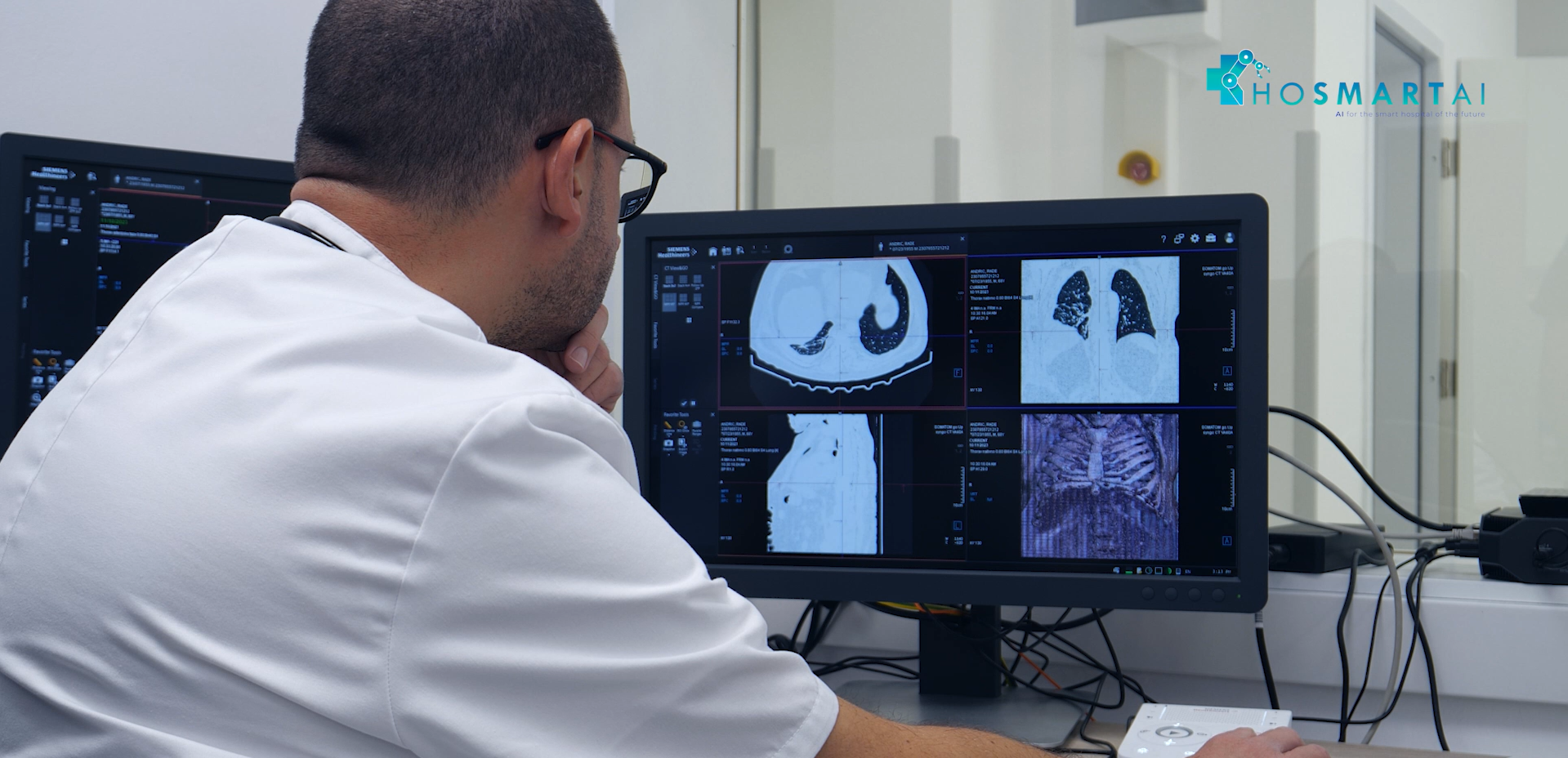
Video: Meet SoftLungX pilot
SoftLungX project was one of the four winners selected for HosmartAI, via Open Call #2: They proposed a Software for Diagnosis of Lung Diseases from Chest X-ray Images.
After successfully passing the two first phases of the programme – Design phase, followed by the Develop & Deploy & Operate phase – they are now on the final stage – Assess. And they have been reaching great achievements!
THE PROBLEM
Lung diseases are some of the most widespread diseases globally. They present a heavy burden on the world healthcare institutions. Over the last couple of years, this burden only increased with the recent Covid-19 pandemic. It is of great importance for lung diseases to be discovered early and treated in a timely manner. Multiple different imaging modalities are used worldwide for diagnosing lung diseases, the most common of which is X-ray due to its low cost, wide availability and ease of use. However, chest X-ray images take a lot of time and human resources to analyse and interpret X ray images. The number of medical professionals available for these tasks stays stagnant, while the number of patents increases over the years. For this reason, it is necessary to create decision support systems capable of helping medical professionals in analysing chest X-ray images, thus shortening the time needed for each examination. Through this endeavour, patient quality of life is increased by reducing the amount of time needed for accurate diagnosis.
THE SOLUTION
SoftLungX offers the solution for these problems through an AI based approach. The system offers an automatic disease classification neural network capable of predicting 17 different lung diseases and radiological findings, with an option of finding multiple lung diseases and radiological findings classes within a single image based on transfer learning. The model is also capable of distinguishing between diseased and healthy lungs. In addition, the model offers a region of interest extraction module, through segmentation with a backbone of U-net, for separating lungs from surrounding tissue within the X-ray image. Segmentation allows for better judgment and decision making from the medical professional using the system. Furthermore, the system offers a gradient class activation map (Grad-CAM) based module for additional decision explainability. The system is capable of making predictions as a support to the medical professionals and providing additional information about the X-ray, reducing the amount of time needed for diagnosis. The Grad-CAM also serves as a way of increasing patient trust in the system.
Pilot video
Check out the video developed by this team, with more details about their innovation, feedback from its users and the expected results and benefits.


