Mapping and ablation of cardiac arrhythmias
Atrial fibrillation is the most common type of heart arrhythmia affecting over 45 million people worldwide. The condition occurs when the upper and lower chambers are not coordinated causing the heart to beat too quickly, slowly, or irregularly. Besides medical treatment, radio-frequency ablation is a minimally invasive catheter-based method using an electrode to destroy or isolate the arrhythmia-triggering tissue.
This pilot project aims to develop surgical tools that assist surgeons in performing the procedure of catheter ablation more effectively. The first tool interprets and visualizes electrophysiology (EP) mapping data for catheter ablation procedures in real-time. The second tool is a robotic system designed to support the navigation of a catheter during catheter ablation using magnetic fields. The robotic system uses the EP mapping algorithm to identify ablation targets and navigates the catheter to the desired targets. With the development and validation of these tools, we work on automating a medical procedure which in future could be conducted by telesurgery. The robotic platform is evaluated in an in-vitro setup by simulating the ablation of tissue around the pulmonary veins, preventing the electrical signals of the veins from disturbing the natural heartbeat. In this setup two cameras are used to track the position of the color markings on the catheter, allowing for feedback control. A GUI shows the surgeon an overview of all the ablation targets and their status. By applying an external magnetic field with an electromagnetic system, the magnetic tip of the catheter can be steered semi-automated or fully automated. In the semi-automated mode, the surgeon controls the magnetic field direction and catheter insertion using a controller interface. The fully automated mode first computes the desired trajectory of the tip and then autonomously navigates the catheter to the targets.
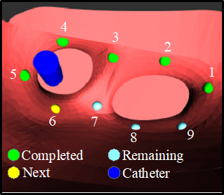
The GUI shows the surgeon the status of the ablation targets and the location of the catheter tip.
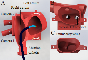
Heart phantom used for the in-vitro experiments. (A) The ablation catheter is advanced from the femoral vein to the right atrium and into the left atrium through a transseptal puncture. (B) The two endoscopic cameras track the position of the catheter tip. (C) The triggers causing atrial fibrillation often lie in the pulmonary veins. In pulmonary vein isolation, the operator ablates points around these veins to electrically isolate the triggers and restore the normal heartbeat.